Product name
Recombinant human PNMA1 protein (tagged)
Purity: > 85% SDS-PAG.

Expression system: Escherichia coli
Accession: Q8ND90
Protein length: full length protein
Animal-free: No
Nature: recombinant
Species: Human
Predicted Molecular Weight: 47 kDa including tags
Amino acids: 1 to 353
Tags: Your N-Terminus Tag
Additional sequence information
N-terminal 10xHis-B2M-JD-tagged and C-terminal Myc-tagged.
Specifications
Our Abpromise warranty covers the use of ab236342 in the following tested applications. Application notes include recommended starting dilutions; the end-user must determine the optimal dilutions/concentrations.
Applications: SDS PAGE
Form: Liquid
Stability and Storage
Shipped at 4°C. Store at -20°C or -80°C. Avoid freeze/thaw cycle.
- pH: 7.2
- Components: Tris buffer, 50% glycerol (glycerin, glycerin)
Abstract
Using spectroscopic, calorimetric, and microscopic methods, we demonstrate that calcium binds to N-terminal 10xHis beta-2-microglobulin (β2M)-JD-tagged Recombinant under physiological conditions of pH and ionic strength, in biological buffers, causing a conformational change associated with the binding of up to four calcium atoms. calcium per β2m molecule, with a marked transformation from some random coil structure to a beta-sheet structure, and culminating in protein aggregation at physiological (serum) concentrations of calcium and β2m. We draw attention to the fact that the β2m sequence contains several potential calcium-binding motifs of the DXD and DXDXD (or DXEXD) varieties.

We establish (a) that the observed microscopic aggregation at physiological concentrations of β2m and calcium converts to actual turbidity and visible precipitation at higher concentrations of protein and β2m, (b) that this initial aggregation/precipitation leads to the formation of amorphous aggregates, (c) that the formation of the amorphous aggregates can be partially reversed by the addition of the divalent ion chelating agent, EDTA, and (d) that after incubation for a few weeks, the amorphous aggregates seem to favour the formation of amyloid aggregates that are They bind to the dye, Thioflavin T (ThT), causing an increase in dye fluorescence.
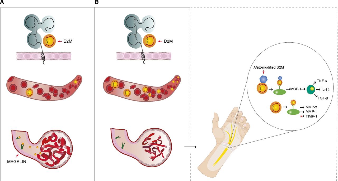
We speculate that β2m exists as microscopic aggregates in vivo and that these do not progress to form larger amyloid aggregates because protein concentrations remain low under normal conditions of renal function and β2m degradation. However, when renal function is compromised, and especially when dialysis is performed, β2m concentrations probably increase transiently to produce large aggregates that are deposited in bony joints and are transformed into amyloid during dialysis-related amyloidosis.

